
Unlocking Human-Relevant 3D Models with VitroScreenORA®
VitroScreenORA® is a proprietary human cell-based, scaffold-free
3D spheroid screening platform, that provides physiologically relevant, predictive, and ethically advanced in vitro models.
3D spheroid screening platform, that provides physiologically relevant, predictive, and ethically advanced in vitro models.
About VitroScreenORA®
VitroScreenORA® spheroids replicate human tissue architecture at the microscale, dynamically evolving through physiological cell-ECM interactions.
These models provide a robust gateway to Microphysiological Systems (MPS) applications, enabling high-precision evaluation of drug accumulation, metabolism, and mechanisms of action through integrative genomics, proteomics, and spatial biology analyses.
VitroScreenORA® Spheroids series at A glance
VitroScreenORA® series
Therapeutic areas
ORA® Series human 3D spheroids
Vascularized and co-culture with immunocompetent cells
Customized system
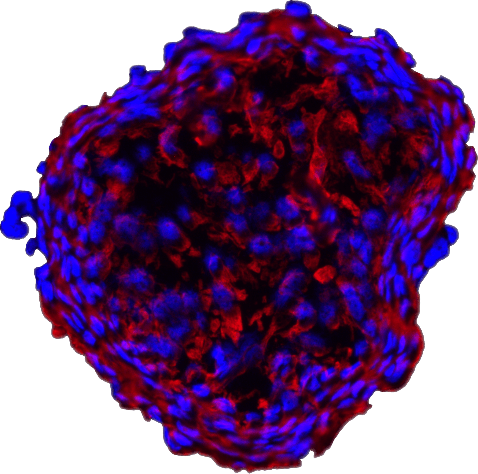
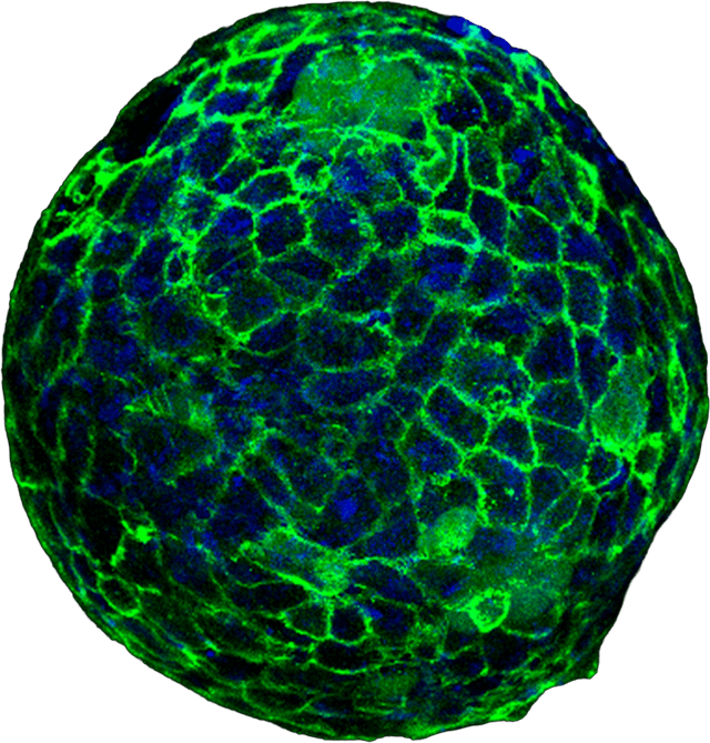
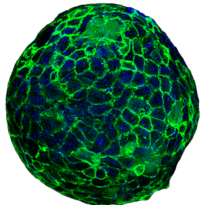
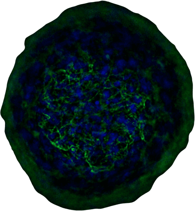
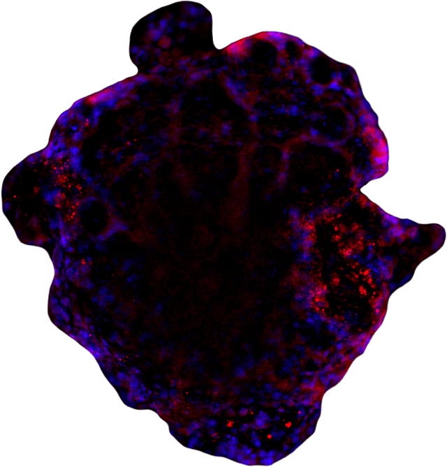
High Content Imaging applied to Spheroids
High Content Imaging enables precise morphological assessment
and biomarker localization within scaffold-free spheroids,
enhancing multi-endpoint analyses in translational research.
Our histo-morphology platform integrates both standard and customized staining and immunofluorescence workflows on paraffin-embedded sections and whole-mount samples, maximizing analytical depth and scientific accuracy.
Drag
Dermal papilla CD31
Cartilage CD44
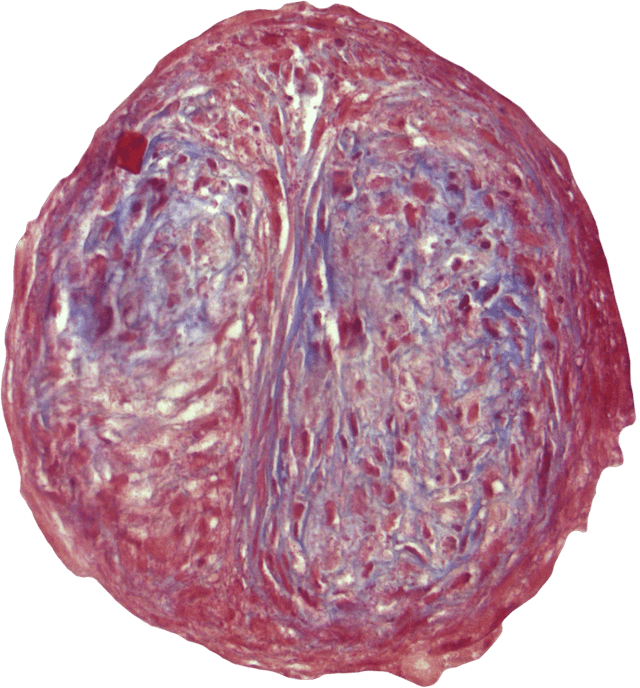
Cornea Masson's Trichrome

Endometrium vim
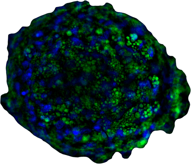
Adipe Nilered
Intestine ZO1
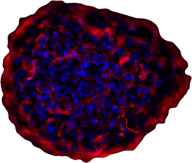
Dermis ColIII
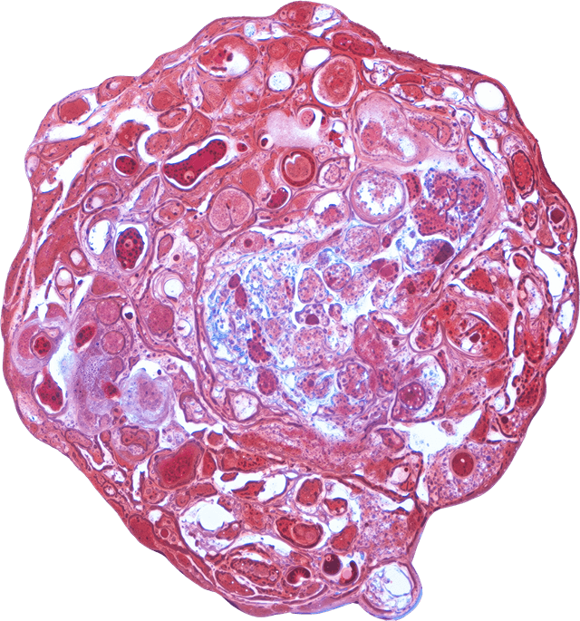
Prostate Masson's Trichrome
Dermal papilla CD31
Cartilage CD44
Cornea Masson's Trichrome
Endometrium vim
Adipe Nilered
Intestine ZO1
Dermis ColIII
Prostate Masson's Trichrome